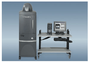
Dedicated equipment and experienced scientists are available to select the best solution to suite your goals.
Imaging is a non-invasive technique that allows repeated measurements of disease progression or recovery in the same subject, which exactly mimics the clinical condition, thus saving animals, time and money. Impressive resolution of anatomical details are obtained even in smallest animals, and functional imaging gives important information on metabolism.
The recent development of genetically-modified animals opens the opportunity to visualize in real time almost any physiological and pathological process in small animals, with substantial boosting of the screening efficacy as well as the understanding of the mechanism(s) of action. The cost-effectiveness of this approach is particularly advantageous during the screening for efficacy.
Magnetic resonance, Nuclear medicine and X-Ray, Ultrasound Imaging and Optical Imaging facilities are available to provide studies for the evaluation of drug candidates in different areas such as oncology, cardiovascular, cerebral, inflammation, drug-absorption and gene expression.


Mice can be genetically modified by inserting a stable luciferase gene to result in an easy-to-detect light signal in response to tissue specific activity. This enables monitoring physiology, pathology, drug or nutraceutical actions, toxic effects in the intact living organism.
Skin Wound Healing assay inexpensive, direct and accurate in vivo quantification of proliferation in wound healing.

